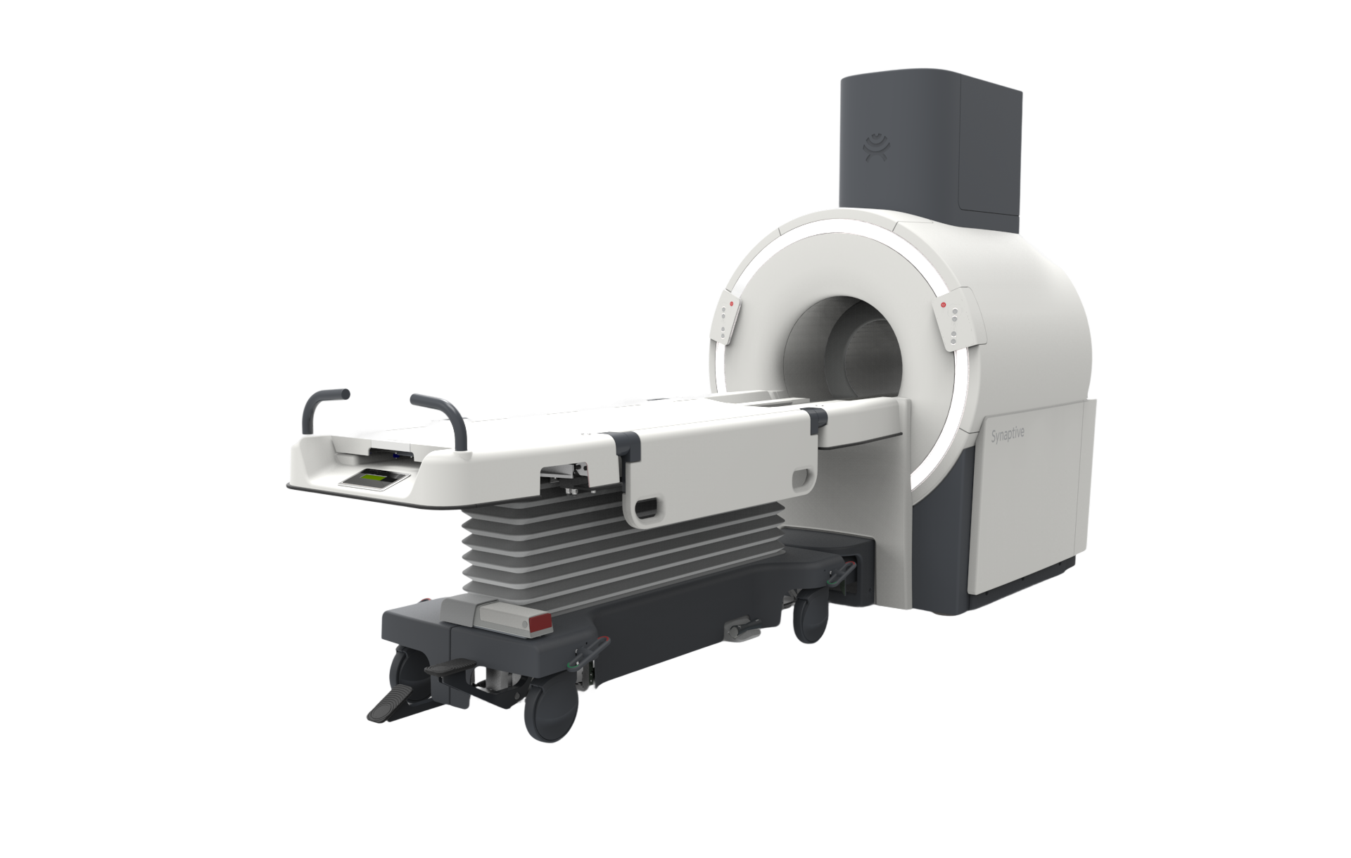
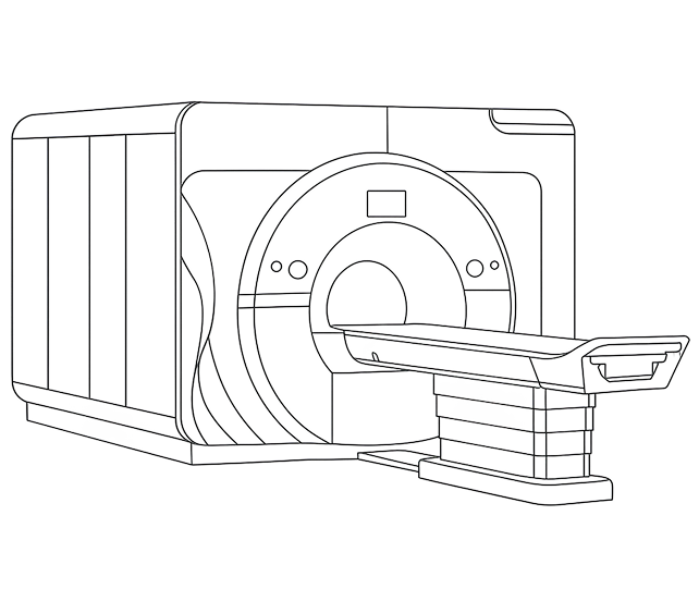
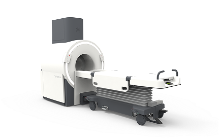
EVRY MRI: The Future of Imaging is Here
A next-generation, low-field 0.5T MRI with best-in-class head imaging
Benefits
The Future of MRI
MRI Features
Full suite of neuro imaging protocols covering broad clinical applications
Unique design with best-in-class geometric accuracy
Footprint 70% smaller than traditional MRIs
No requirement for helium cooling and venting
Clinical/Economic Benefits
Qualifies for same high-field MRI reimbursement codes
High precision targeting for MRI-guided interventions
Permits increased access to MRI in every area of the hospital
Highly adaptable for low-and-middle-income regions
Comparison
EVRY MRI vs. Legacy MRI
EVRY MRI vs. Legacy MRI
Comparison
EVRY
Legacy
Install
8x–40x Lower Install Costs
-
Space
250 sq. ft.
1,000+ sq.ft
Weight
4,000 lbs
20,000 lbs
Safety
70% Less Energy with on/off flexibility
Always on
Geometric Distortion (max)
0.5mm
5mm
Acoustic Noise
No hearing protection required
-
EVRY EXCEEDS LEGACY MRI PERFORMANCE WITH 1/5 THE FOOTPRINT AND COST
EVRY outperforms legacy MRI - opening access, lowering cost, and powering precision therapies.
3D T1
1.5T
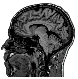
3D MPRAGE
1.0 x 1.0 x 1.0mm
5:45
1.0 x 1.0 x 1.0mm
5:45
0.5T
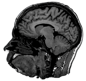
3D SPGR
1.1 x 1.1 x 1.1mm
5:30
1.1 x 1.1 x 1.1mm
5:30
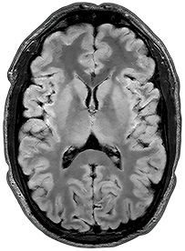
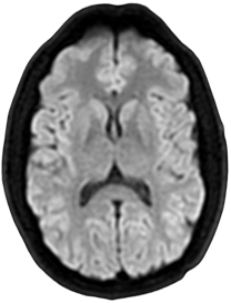
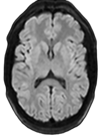
3D FLAIR
1.5T
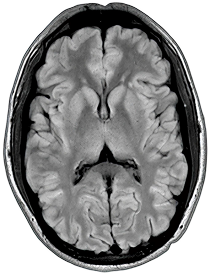
1 x 1 x 3mm
3:31
3:31
0.5T
1 x 1 x 3mm
3:15
3:15
DWI
1.5T
2 x 2 x 5mm
1:02
1:02
0.5T
2 x 2 x 5mm
1:35
1:35
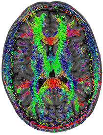
DTI - Tractography
1.5T
3 x 3 x 3mm
5:10
5:10
0.5T
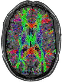
3 x 3 x 3mm
10:25
10:25
3D FLAIR
Tumor (T1+c)

1.5T
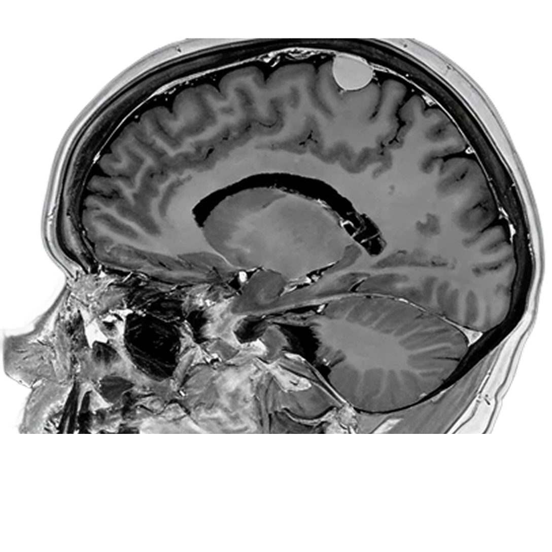
0.5T
fMRI
Functional (fMRI)
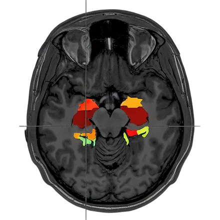
3.0T
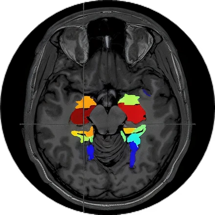
0.5T
Images from: Halder et al., Investigating the feasibility of resting state functional MRI with GRE EPI on a high performance 0.5 T Scanner, processed using GraphICA, an asset of Brainet-Brain Imaging Solutions Inc.
Engineered for the Brain Computer Interface Era
Built for the Future
Legacy MRI technologies were not built to address the complexities of imaging patients with implants.
3T
LEGACY
Imaging at higher field inherently increases the risk of implant heating and associated tissue damage.

0.5t
EVRY

Lower field strength MRI reduces the risk of implant heating and associated tissue damage.
Patients with implants often face imaging restrictions due to the risk of implant heating and associated tissue damage.
With its unique design, the EVRY system minimizes exposure to electric and magnetic fields, opening the door to more imaging possibilities for patients with implants.