Why Tractography

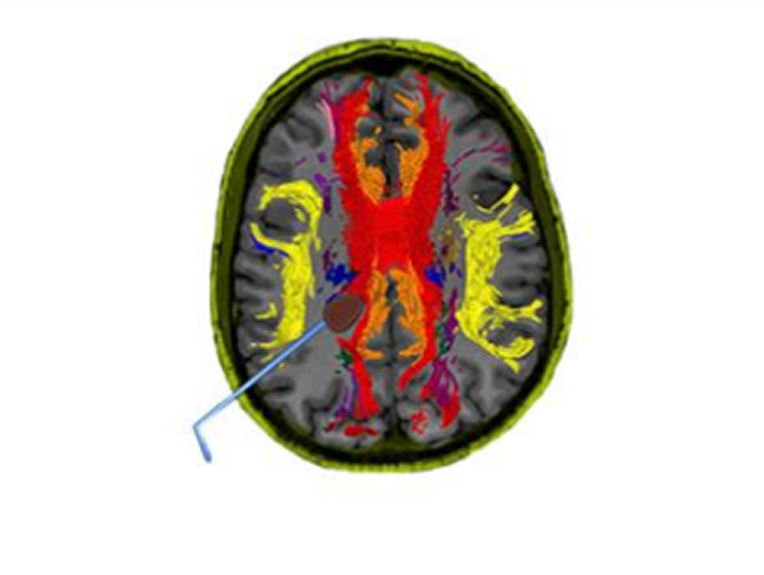
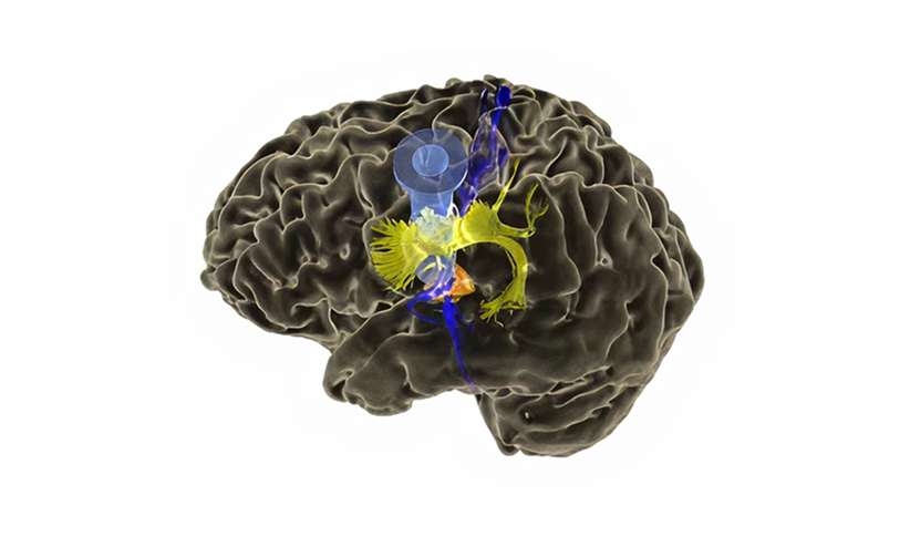
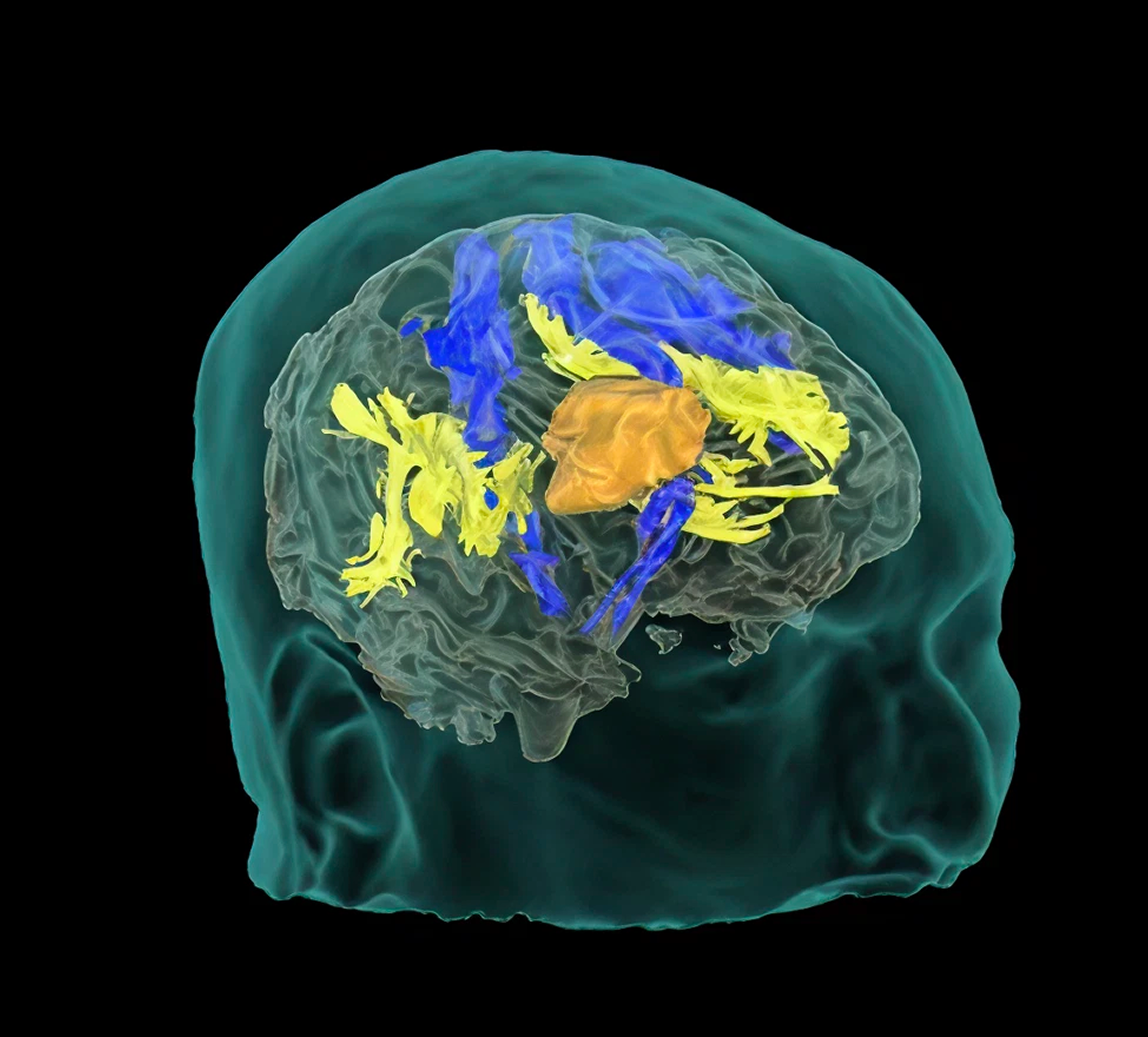
Tractography is a non-invasive tool that infers the location of the white matter. Understanding this patient-specific information may be used to inform safer treatment.

Tractography can be used to determine an approach to a tumor, to help define boundaries of resection, or for postoperative tracking of patient recovery.

Modus Nav Overview

Colored brain scan highlighting neural pathways in red, yellow, and orange on a gray background.

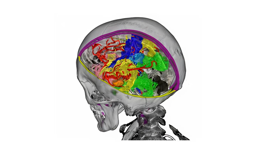
Leverage brain mapping to support safe maximal resection

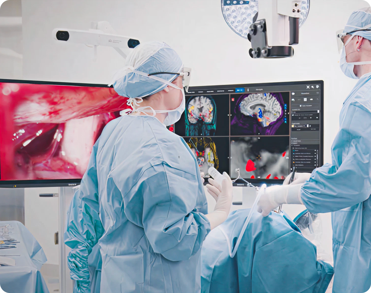
Navigate intraoperative tractography with automated white matter segmentations unaffected by pathology-induced edema.

3D illustration of a human brain with highlighted internal structures in yellow, blue, and orange.

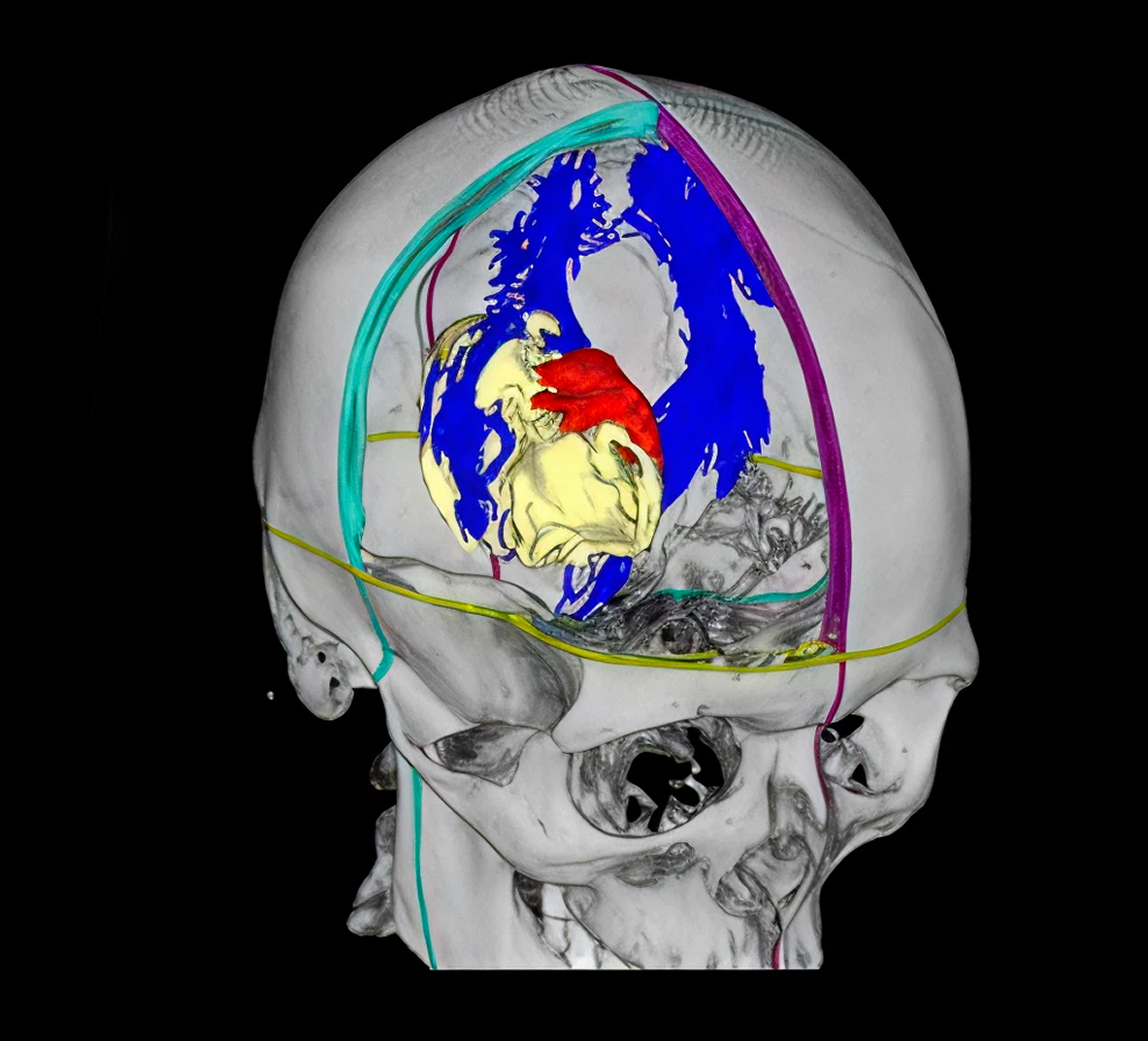
Accurately track multiple intraoperative instruments

Instrument position and trajectory are accurately navigated and displayed in real time - including suction tools and MIS ports.

3D medical scan showing a semi-transparent skull with colorful brain structures and blood vessels inside.

Precision medicine workflows through seamless integrations

Modus Nav combines data elements from Modus Plan, combining critical pre-surgical imaging data with Modus X optics in the OR.

MODUS NAV

How Surgeons Are Using Modus Nav

Watch Video Now

Tumor Resection

3D medical illustration of a semi-transparent human head with brain structures highlighted, including yellow, blue, and orange regions indicating neural pathways or tumor areas.

AVM Resection

3D anatomical visualization of a human skull with highlighted brain regions in blue, a red area, and a yellow structure indicating the heart inside the skull.

ICH Evacuation

3D brain scan showing internal structures highlighted in yellow, blue, and orange inside a translucent skull.

Functional Targeting

3D brain scan showing colored neural pathways and brain structures in a child's head profile.

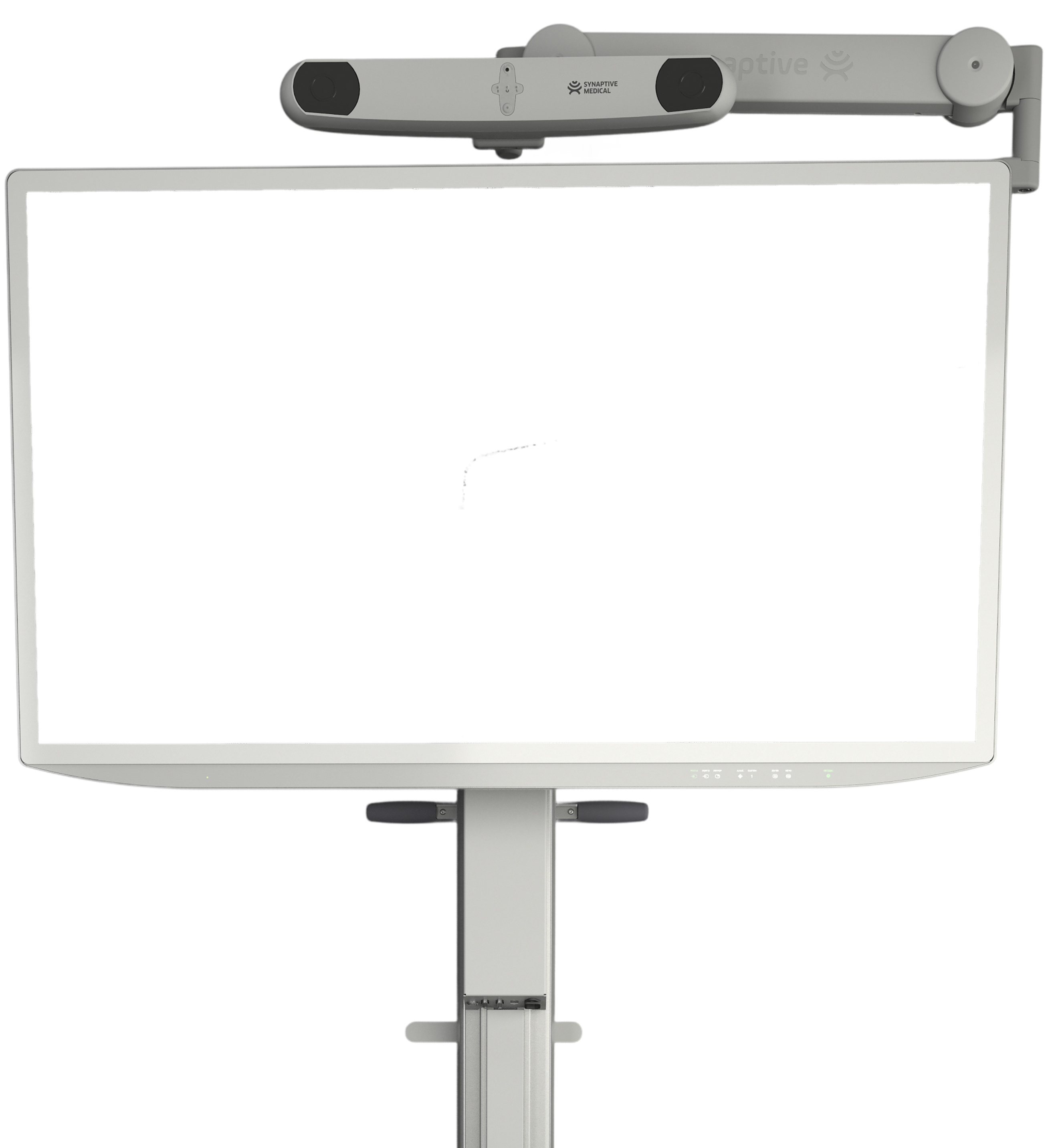
Modus Nav and Modus X: Seamless Integration of Visualization and Navigation

Ensure consistency and accuracy of the surgical approach by integrating Modus Nav with Modus X, Synaptive's 3D robotic exoscope. This ecosystem of products come together to create a modern neurosurgical experience delivering novel patient information with automated efficiency.

Two surgeons in blue scrubs and masks performing brain surgery with brain scans displayed on monitors in the operating room.

What Surgeons Say

Webinar announcement for a panel discussion on clinical use of tractography in neurosurgery with photos of three doctors.

Panel Discussion on the Clinical Use of Tractography in Neurosurgery

Dr. Chaichana (Mayo Clinic), Dr. Schulder (Northwell Health) and Dr. Dickson (Sutter Health Eden Medical Center) discuss the importance of white matter and their experience using tractography in neurosurgery.

Watch Video Now
Webinar announcement for a case review using the Robotic Digital Microscope Modus V in brain and spine surgery on Monday, May 3rd at 12:00 PM and 8:00 PM EDT with Dr. Peter Fecci from Duke Health, Dr. Costas Hadjipanayis from Mount Sinai Hospital, and Dr. Raj Nangunoori from Mercy Health.

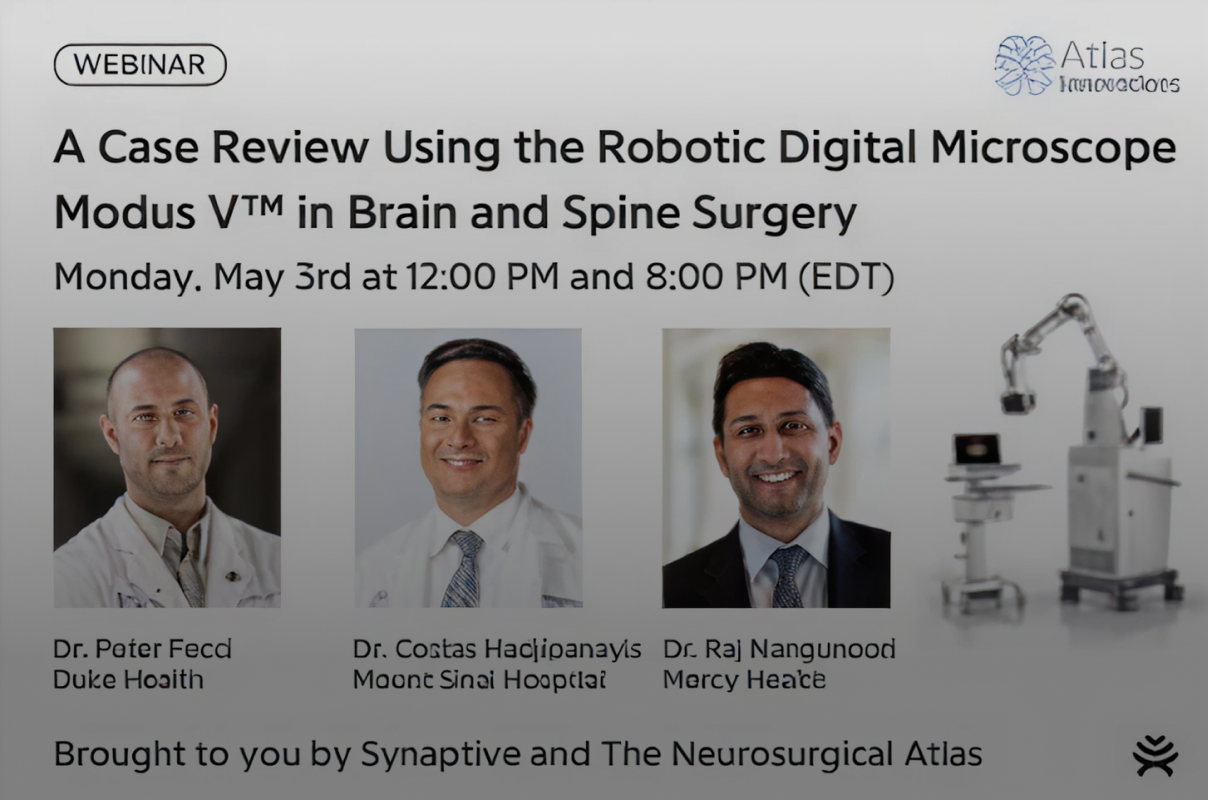
A Case Review Using the Robotic Digital Microscope Modus V in Brain and Spine Surgery

In partnership with The Neurosurgical Atlas, Dr. Peter Fecci (Duke Health), Dr. Costas Hadjipanayis (Mount Sinai, NYC) and Dr. Raj Nangunoori (Mercy Health) present on their experience using Synaptive's surgical products in cranial and spine surgery.

Watch Video Now
Presentation slide titled 'Clinical Applications of Tractography and a Preview of a Novel Automatic Segmentation Algorithm' by Dr. Sebastian Koga from Ochsner Health System, with an image of a man speaking.

Clinical Applications of Tractography and a Preview of a Novel Automatic Segmentation Algorithm

Dr. Sebastian Koga from Ochsner Health System in Louisiana speaks about the clinical applications of tractography in his surgical practice, as well as beyond surgery, for use in longitudinal tracking of post-operative patient recovery.

Watch Video Now

In The Literature

Frontiers in Neurology journal cover titled 'Preoperative validation of edema-corrected tractography in neurosurgical practice: translating surgeon insights into novel software implementation' featuring brain scan images with colorful tractography overlays.
Koga et al., 2024

Preoperative validation of edema-corrected tractography in neurosurgical practice: translating surgeon...

See All Research
Seal of Cureus Journal of Medical Science with caduceus symbol and established year 2009.
Zakaria et al., Cureus, 2017

Automated Whole Brain Tractography Affects Preoperative Surgical Decision Making

See All Research
Circular teal emblem with text 'Cureus Journal of Medical Science' around the edge, 'EST. 2009' and a medical caduceus symbol inside.
Agarwal et al., Cureus, 2017

Tractography for Optic Radiation Preservation in Transcortical Approaches to Intracerebral...

See All Research