MODUS PLAN

Dynamic Free Water Correction

MRI brain scan with colorful diffusion tensor imaging showing neural fiber tracts and a visible abnormal lesion on the right hemisphere.
Uncorrected
Axial brain MRI scan with colorful fiber tractography showing neural pathways in red, green, and blue.
Corrected

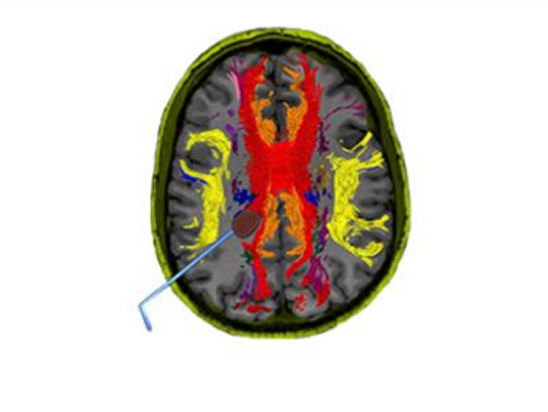
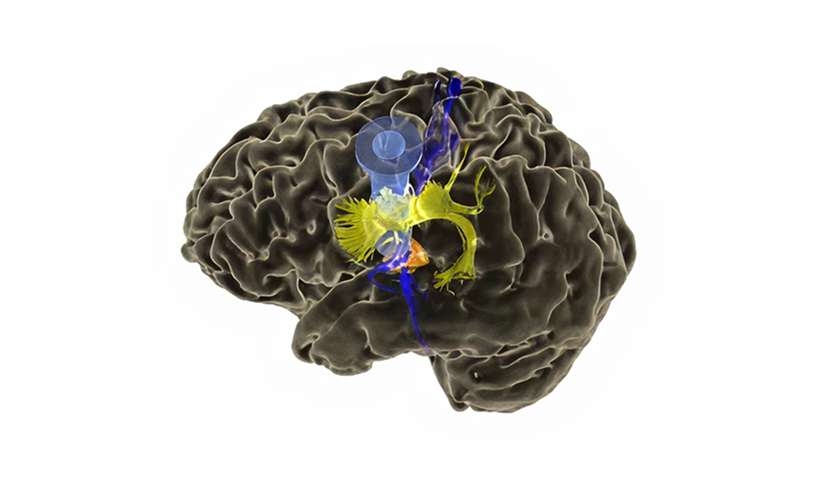
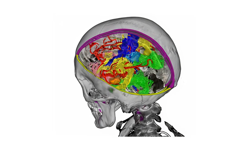
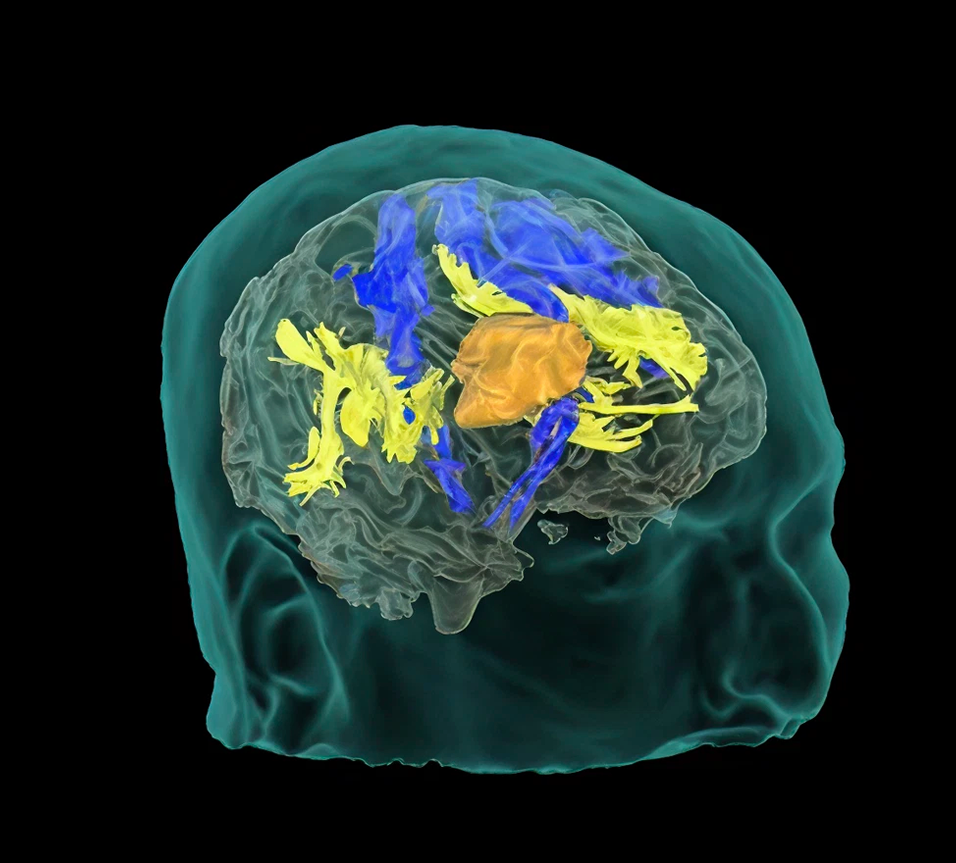
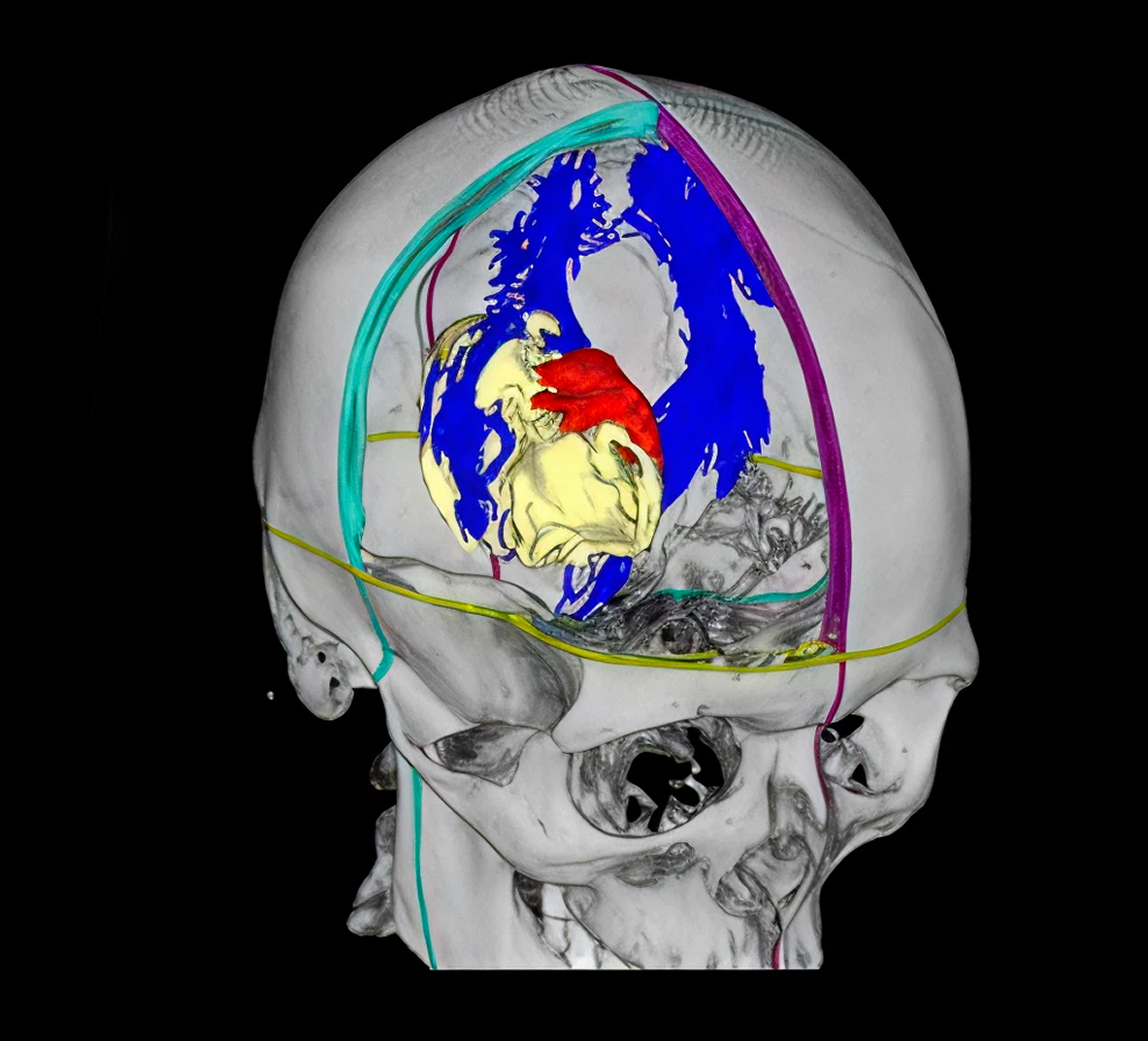
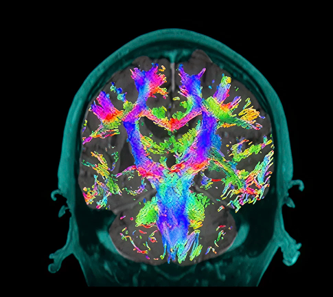
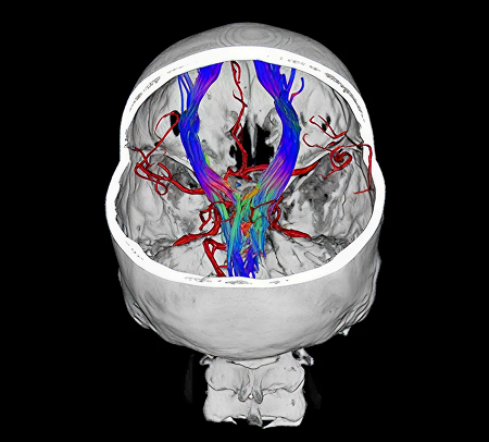
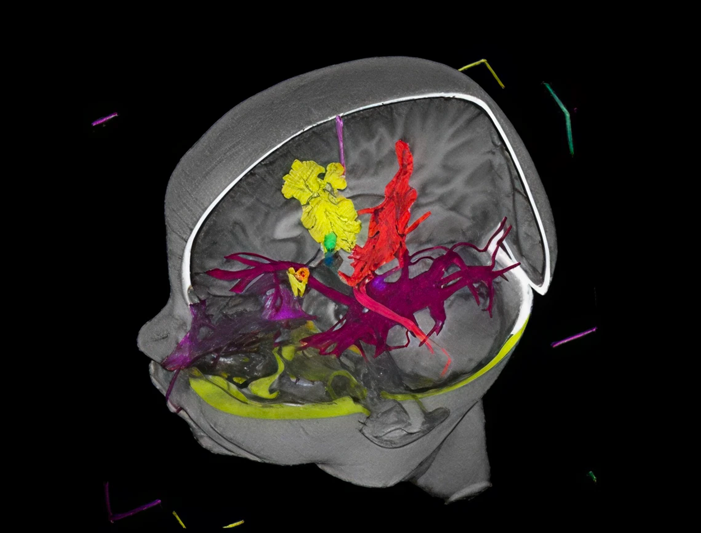
Synaptive's Dynamic Free Water Correction software automatically applies a correction to tractography that allows for the recovery of eloquent tracts in areas around brain lesions previously impacted by edema. This information can be combined with vascular, bone and functional imaging, allowing users to see their patients in new ways to offer better treatment.

See Reveal For Modus Plan
Why Tractography

Tractography is a non-invasive tool that infers the location of the white matter. Understanding this patient-specific information may be used to inform safer treatment.

Tractography can be used to determine an approach to a tumor, to help define boundaries of resection, or for postoperative tracking of patient recovery.

Modus Nav Overview

Colored brain scan highlighting neural pathways in red, yellow, and orange on a gray background.

Easy access to tractography

Synaptive automatically groups tracts into patient-specific bundles of interest such as the corticospinal tract or arcuate fasciculus, making it easy for the surgical team to access tractography when and where they need it - for every patient.

Watch Video Now
3D illustration of a human brain with highlighted internal structures in yellow, blue, and orange.

Enables precision medicine to support value-based care

Modus Plan Allows surgeons to perform immersive simulations of various minimally invasive surgical approaches, and tailor their treatment to that individual patient.

Watch Video Now
3D medical scan showing a semi-transparent skull with colorful brain structures and blood vessels inside.

Dynamic free water correction

Software automatically applies a correction to tractography that highlights tracts in areas that would ordinarily be obscured by diffusion signal wash out, such as areas of edema.

Watch Video Now
Modus Plan

How Surgeons Are Using Modus Plan

See The Clinical Use of Tractography

Tumor Resection

3D medical illustration of a semi-transparent human head with brain structures highlighted, including yellow, blue, and orange regions indicating neural pathways or tumor areas.

ICH Evacuation

3D brain scan showing internal structures highlighted in yellow, blue, and orange inside a translucent skull.

AVM Resection

3D anatomical visualization of a human skull with highlighted brain regions in blue, a red area, and a yellow structure indicating the heart inside the skull.

Post-op Assessment

Coronal brain MRI scan with colorful fiber tractography showing neural pathways.

Artery Bypass

3D visualization of the human skull showing colored brain blood vessels and neural pathways inside.

Functional Targeting

3D brain scan showing colored neural pathways and brain structures in a child's head profile.

Modus Plan Purchase Options

Purchase Modus Plan

Purchase Modus Plan as a standalone product or as part of a Synaptive product bundle.

Rent-to-Own Modus Plan

This rent-to-own option includes flexibility to build toward ownership.

Modus Plan On-Demand

This program allows surgeons quick, on-demand access to patient-specific tractography when they need it. Includes building toward ownership.

What Surgeons Say

Webinar announcement for a panel discussion on clinical use of tractography in neurosurgery with photos of three doctors.

Panel Discussion on the Clinical Use of Tractography in Neurosurgery

Dr. Chaichana (Mayo Clinic), Dr. Schulder (Northwell Health) and Dr. Dickson (Sutter Health Eden Medical Center) discuss the importance of white matter and their experience using tractography in neurosurgery.

Watch Video Now
Webinar announcement for a case review using the Robotic Digital Microscope Modus V in brain and spine surgery on Monday, May 3rd at 12:00 PM and 8:00 PM EDT with Dr. Peter Fecci from Duke Health, Dr. Costas Hadjipanayis from Mount Sinai Hospital, and Dr. Raj Nangunoori from Mercy Health.

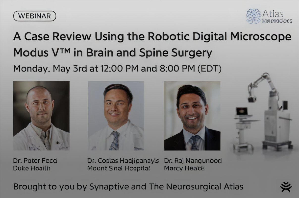
A Case Review Using the Robotic Digital Microscope Modus V in Brain and Spine Surgery

In partnership with The Neurosurgical Atlas, Dr. Peter Fecci (Duke Health), Dr. Costas Hadjipanayis (Mount Sinai, NYC) and Dr. Raj Nangunoori (Mercy Health) present on their experience using Synaptive's surgical products in cranial and spine surgery.

Watch Video Now
Presentation slide titled 'Clinical Applications of Tractography and a Preview of a Novel Automatic Segmentation Algorithm' by Dr. Sebastian Koga from Ochsner Health System, with an image of a man speaking.

Clinical Applications of Tractography and a Preview of a Novel Automatic Segmentation Algorithm

Dr. Sebastian Koga from Ochsner Health System in Louisiana speaks about the clinical applications of tractography in his surgical practice, as well as beyond surgery, for use in longitudinal tracking of post-operative patient recovery.

Watch Video Now
Presentation slide titled 'Oncologic and Neurovascular Case Experience with Synaptive's Modus V and Integrated Tractography Platform' featuring a photo of a man in a suit standing in a medical setting.

Oncologic and neurovascular case experience with Modus V and integrated tractography platform

Dr. Lawrence Dickinson from Sutter Health Eden Medical Center in California speaks about the advantages of Modus V and tractography in his surgical practice. He also shares tips on how to set up а business plan.

Watch Video Now

See How Modus Plan Has Impacted Real Patients

Smiling woman holding torn photos with outdoor scenes in both hands.

Tiffany's story

Tiffany's motor function was preserved during brain tumor surgery.

Middle-aged man wearing a black cap, red shirt, and black jacket sitting outdoors near a body of water with houses and trees in the blurred background.

David's story

The Synaptive solution contributed to making an inoperable tumor operable.

In The Literature

Title text reading 'WORLD NEUROSURGERY' in large, gradient gold and white letters on a black background.
February 2024

Transcorticosubcortical Approach for Left Posterior Mediobasal Temporal Region Gliomas: A...

See All Research
Logo for Operative Neurosurgery with a stylized neuron symbol inside the letter O.
January 2024

The Surgical White Matter Chassis: A Practical 3-Dimensional Atlas for...

See All Research
Seal of Cureus Journal of Medical Science with caduceus symbol and established year 2009.
January 2024

Automated Whole Brain Tractography Affects Preoperative Surgical Decision Making

See All Research

Integration Across the Synaptive Integrated Suite Allows You to:

Automatically quality-control your data

Synaptive's ImageDrive™ Clinical ensures data is of sufficient quality for clinical use-in this case, to generate tractography. Users will get updates in real time as to whether data is suitable for its intended use, so surgeons get the right data when they need it.

Sync your plans and data across Synaptive systems

ImageDrive Clinical is a hub between your Synaptive products. With ImageDrive Clinical, data is transferred seamlessly between planning and navigation stations for easy collaboration. Multi-modal data is stored chronologically for longitudinal review, including DICOM, pre- and post-op tractography, Modus V video and navigation captures.

Visualize dynamic tractography and surgical plans in the OR

Send tractography and surgical plans to Synaptive's navigation, BrightMatterT™ Guide, for seamless and easy access to dynamic tractography in the OR. Surgeons can then automatically align Modus V to a pre-op or intra-op trajectory, enabling hands-free visualization of the operative field.

Contact us to Learn More Imagedrive Clinical and Navigation